


When breast cancer is diagnosed in its earliest stages, the survival rate is nearly 100%. However, for tumours detected in later stages, that rate drops significantly to around 25%.
In a quest to enhance the overall survival rate for breast cancer patients, researchers from MIT have developed a wearable ultrasound device. This technology could enable individuals to detect tumours in their early stages, which is crucial for improving outcomes. The device proves particularly valuable for patients with a high risk of developing breast cancer in the time between routine mammograms.
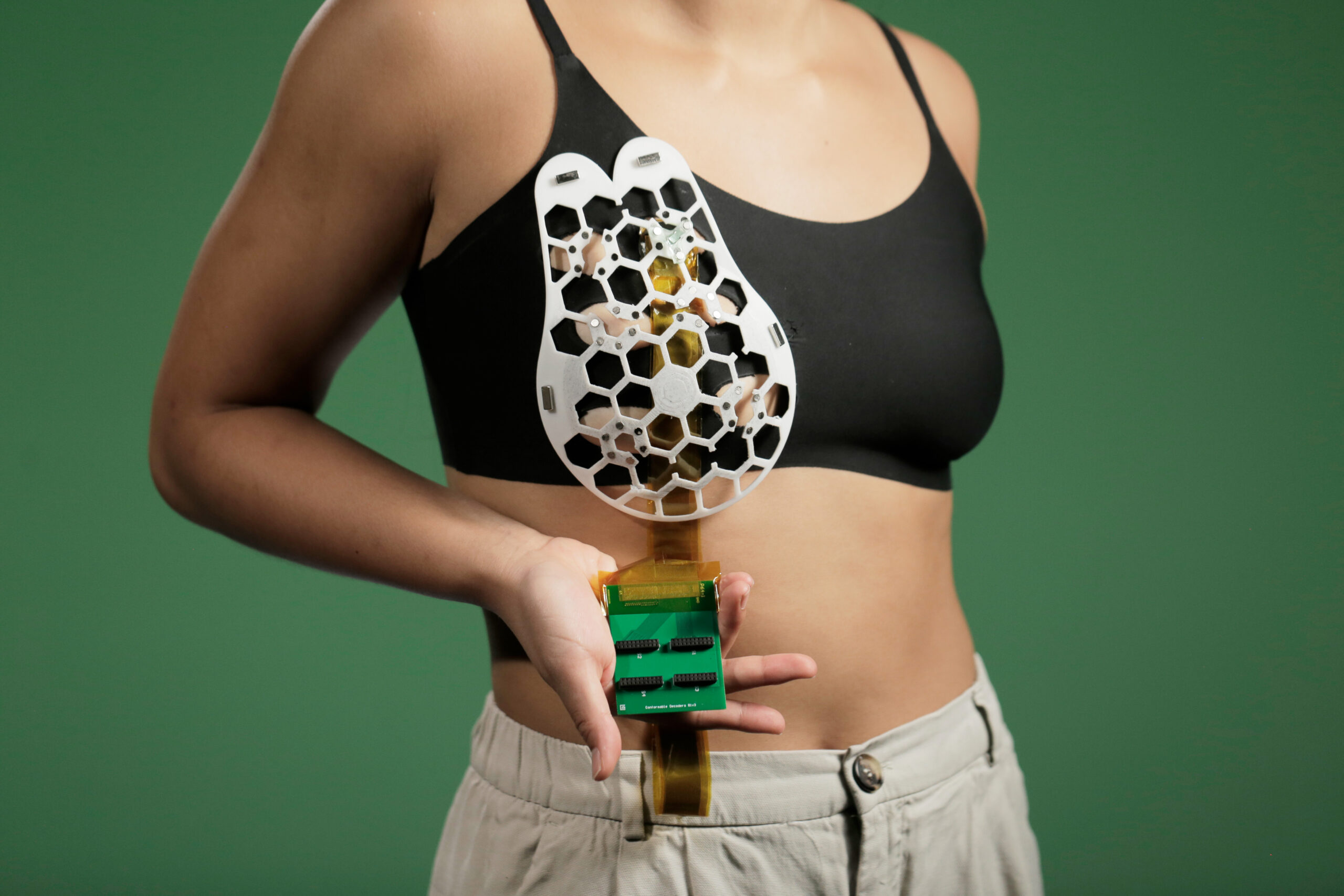
The device is a flexible patch that can be conveniently attached to a bra, providing the wearer with the ability to move an ultrasound tracker along the patch and image the breast tissue from various angles. In a recent study, the researchers demonstrated that they were able to obtain ultrasound images with resolution comparable to those produced by the ultrasound probes used in medical imaging centres.
“We changed the form factor of the ultrasound technology so that it can be used in your home. It’s portable and easy to use, and provides real-time, user-friendly monitoring of breast tissue,” says Canan Dagdeviren, an associate professor in MIT’s Media Lab and the senior author of the study.
MIT graduate student Wenya Du, Research Scientist Lin Zhang, Emma Suh ’23, and Dabin Lin, a professor at Xi’an Technological University, are the lead authors of the paper, which appears today in Science Advances.
For this project, Dagdeviren found her inspiration from her late aunt, Fatma Caliskanoglu, who was diagnosed with late-stage breast cancer at the age of 49, despite undergoing regular cancer screenings. Tragically, she passed away only six months later. While at her aunt’s bedside, Dagdeviren, who was a postdoc at MIT at the time, sketched a rough schematic of a diagnostic device that could be seamlessly integrated into a bra. This device would enable more frequent screenings for individuals at high risk of breast cancer, aiming to prevent such devastating outcomes in the future.
Breast tumours that develop in the time between regularly scheduled mammograms, known as interval cancers, constitute approximately 20 to 30% of all breast cancer cases. These tumours often exhibit a more aggressive nature compared to those detected during routine scans.
“My goal is to target the people who are most likely to develop interval cancer,” says Dagdeviren, whose research group specialises in developing wearable electronic devices that conform to the body. “With more frequent screening, our goal to increase the survival rate to up to 98%.”
To turn her vision of a diagnostic bra into reality, Dagdeviren designed a miniaturised ultrasound scanner that enables users to perform breast imaging at their convenience. The scanner is built on the same ultrasound technology used in medical imaging centres, but it incorporates a novel piezoelectric material, which is responsible for the miniaturisation of the ultrasound scanner.
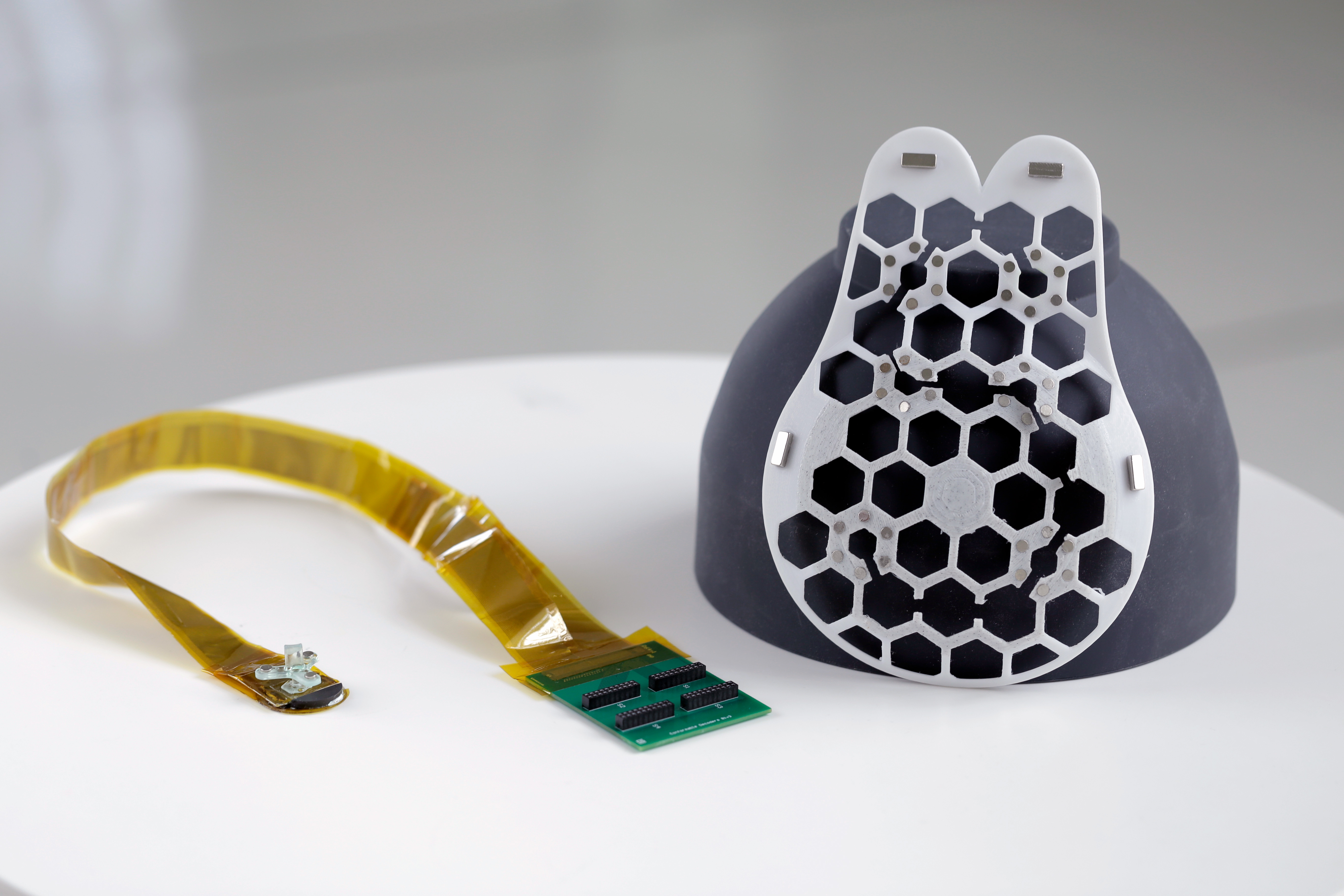
To make the device wearable and user-friendly, the researchers developed a flexible, 3D printed patch featuring honeycomb-like openings. This patch can be easily attached to a bra using magnets, and it aligns with corresponding openings in the bra to allow the ultrasound scanner to make direct contact with the skin. The ultrasound scanner is housed inside a small tracker that can be easily moved to six different positions, ensuring comprehensive imaging of the entire breast.
Furthermore, the scanner can be rotated to capture images from different angles, enhancing its imaging capabilities. The best part is that operating the device does not require any specialised expertise.
“This technology provides a fundamental capability in the detection and early diagnosis of breast cancer, which is key to a positive outcome,” says Anantha Chandrakasan, dean of MIT’s School of Engineering, the Vannevar Bush Professor of Electrical Engineering and Computer Science, and one of the authors of the study. “This work will significantly advance ultrasound research and medical device designs, leveraging advances in materials, low-power circuits, AI algorithms, and biomedical systems.”
Collaborating with the MIT Centre for Clinical and Translational Research, the researchers conducted a test of their device on a single human subject, a 71-year-old woman with a history of breast cysts. Utilising the new wearable ultrasound device, they successfully detected the presence of cysts, some as small as 0.3 centimetres in diameter, which is comparable in size to early-stage tumours. Additionally, the team demonstrated that the device’s resolution matched that of conventional ultrasound technology.
Another noteworthy achievement was the ability to image breast tissue at a depth of up to 8 centimetres, highlighting the device’s capacity to penetrate deeper layers for comprehensive screenings.
“Access to quality and affordable health care is essential for early detection and diagnosis. As a nurse I have witnessed the negative outcomes of a delayed diagnosis. This technology holds the promise of breaking down the many barriers for early breast cancer detection by providing a more reliable, comfortable, and less intimidating diagnostic,” says Catherine Ricciardi, nurse director at MIT’s Centre for Clinical and Translational Research and an author of the study.
At present, to view the ultrasound images, the researchers need to connect their wearable ultrasound scanner to the same type of ultrasound machine typically used in medical imaging centres. However, they are actively developing a miniaturised version of the imaging system, aiming to create a device that is about the size of a smartphone. This innovation will enable users to directly visualise the ultrasound images on the handheld device, eliminating the need for additional connections to external machines.
The wearable ultrasound patch has the advantage of being reusable, making it a cost-effective and practical solution for regular screenings. The researchers have a vision for its application in homes, particularly for individuals at high risk of breast cancer who would benefit from more frequent monitoring.
Furthermore, the device’s portability and ease of use could prove to be a game-changer for individuals who lack regular access to traditional screening facilities. It opens up the possibility of reaching underserved populations and communities with limited healthcare resources, enabling earlier diagnosis and intervention for those who might otherwise go undiagnosed.
“Breast cancer is the most common cancer among women, and it is treatable when detected early,” says Tolga Ozmen, a breast cancer surgeon at Massachusetts General Hospital who is also an author of the study. “One of the main obstacles in imaging and early detection is the commute that the women have to make to an imaging centre. This conformable ultrasound patch is a highly promising technology as it eliminates the need for women to travel to an imaging centre.”
The researchers have a promising vision for the future development of their technology. They aim to create a workflow that utilises artificial intelligence (AI) to analyse the gathered data and track how the ultrasound images change over time. This approach could potentially lead to more accurate and reliable diagnostics compared to the traditional method of relying solely on the subjective assessment of a radiologist comparing images taken years apart.
Additionally, the researchers plan to explore the adaptation of their ultrasound technology for scanning other parts of the body. It could lead to the development of similar wearable devices for monitoring and diagnosing conditions in different organs and body regions, potentially revolutionising healthcare practices and enhancing early detection and treatment for a range of diseases.