


Explore how to overcome these challenges effectively and their popular use cases. Also learn about a 5MP high-sensitive camera by e-con Systems – tailor-made for microscopic-based diagnostic devices.
Microscopic-based diagnostic devices have revolutionised medical diagnosis, enabling detailed examination of cellular and tissue samples. Cameras integrated into these devices are critical in visualisation, documentation, and analysis. However, various imaging challenges exist in different medical segments.
In this blog, e-con System’s explore the major use cases, their challenges, and the types of cameras required to address them effectively. As well as the See3CAM_50CUG – a 5MP high-sensitive camera by e-con Systems that can amplify the performance of microscopic-based diagnostic devices.
Role of cameras in microscopic-based diagnostic devices
Cameras have emerged as indispensable tools in advancing microscopic-based medical diagnosis. With their high-resolution sensors and advanced imaging capabilities, cameras enable healthcare professionals to capture and analyse detailed microscopic images with unprecedented clarity. These images serve as valuable visual evidence, aiding in the accurate identification and characterisation of cellular structures, tissue samples, and pathogens.
Furthermore, real-time visualisation and image analysis facilitated by cameras empower medical practitioners to make informed decisions, improve diagnostic accuracy, and enhance patient care.
Major uses cases of microscopic-based diagnostic devices
Histopathology
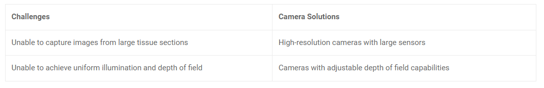
Histopathology is a field that plays a critical role in diagnosing diseases and understanding tissue abnormalities. It involves the examination of tissue samples under a microscope to identify and analyse the changes occurring at a cellular level. By studying the microscopic features of tissues, histopathologists can provide valuable insights into the nature and progression of diseases.
However, histopathology imaging comes with its own set of challenges. The good news is that there are camera solutions to overcome them:
Hematology
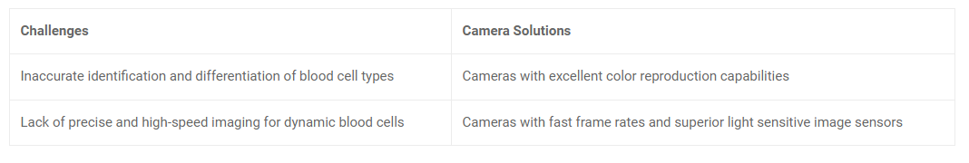
Hematology is a branch of medical science that focuses on studying blood and blood disorders. It involves examining and analysing blood samples to diagnose and monitor various conditions related to blood cells, such as anemia, leukemia, and clotting disorders. In hematology imaging, specific challenges need to be addressed to ensure accurate identification and differentiation of blood cell types. Here is the list of challenges and their corresponding solutions:
Microbiology
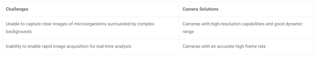
Microbiology is a scientific discipline that focuses on studying microorganisms, including bacteria, viruses, fungi, and other microscopic organisms. It plays a crucial role in various fields, such as medicine, agriculture, environmental science, and biotechnology. Microbiology imaging involves visualising and analysing microorganisms to understand their structure, behaviour, and interactions. However, certain imaging challenges must be addressed to capture clear images of microorganisms, often surrounded by complex backgrounds:
Cytology
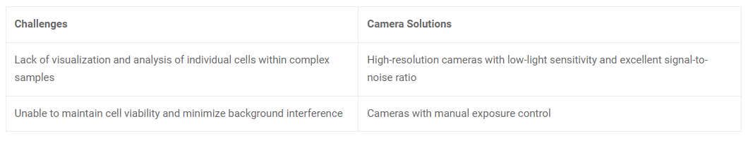
Cytology is a branch of biology that focuses on the study of individual cells to understand their structure, function, and behaviour. In the medical field, cytology plays a crucial role in diagnosing diseases, particularly cancer, by examining cells for abnormalities and identifying malignant or precancerous conditions. Let’s explore their challenges and related camera solutions.
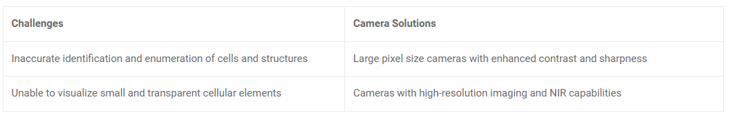
Urine sediment analysis
Urine sediment analysis is a diagnostic procedure involving the microscopic examination of cellular elements and structures in urine samples. It is an essential component of urinalysis that helps assess kidney function, detects urinary tract infections, and identifies various renal disorders. During urine sediment analysis, specific challenges need to be addressed to ensure accurate identification and enumeration of cells and structures:
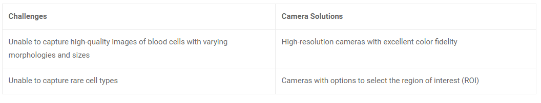
Peripheral blood smear
Peripheral blood smear analysis is a diagnostic technique that examines a thin layer of blood cells spread on a glass slide. It is a valuable tool in haematological investigations and allows for evaluating various blood cell types, their morphology, and any abnormalities present. Here’s a summary of the camera solutions for the challenges faced in peripheral blood smear imaging:
See3CAM_50CUG – a best-fit camera solution by e-con Systems
e-con Systems, a pioneer in the embedded vision space since 2003, offers a wide range of cameras to build medical devices, including diagnostic microscopes.
The See3CAM_50CUG proves to be the ideal camera choice to address the imaging challenges of histopathology, haematology, microbiology, cytology, urine sediment analysis, and peripheral blood smears.
This 5MP high-sensitive colour camera leverages the Sony Pregius IMX264 CMOS sensor for exceptional image quality and accuracy and comes with relevant features such as:
- High-resolution imaging
- Adjustable depth of field
- Excellent colour reproduction
- Advanced image processing capabilities