


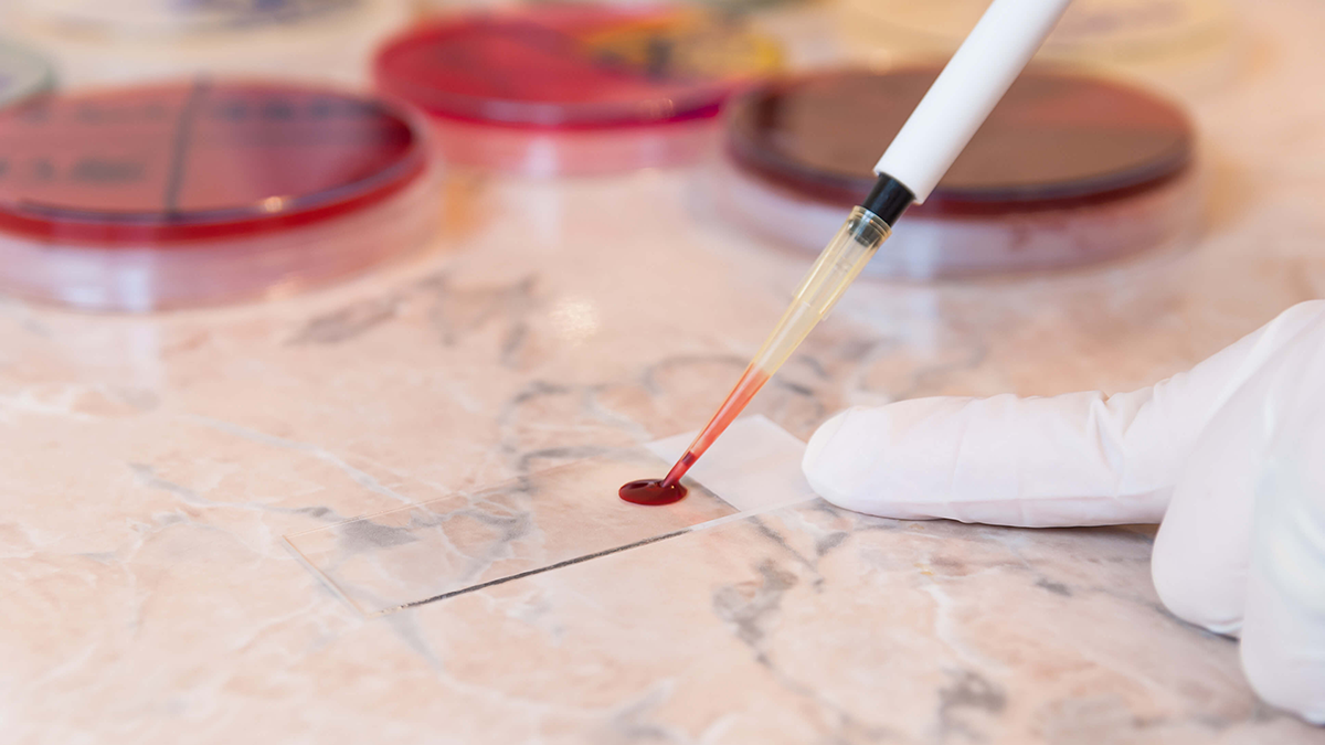
Typically recommended as part of a routine complete blood count test, it offers valuable insights into blood composition. In this piece, we delve into the blood smear testing process and the importance of integrated camera solutions in enhancing its efficacy.
Blood smear testing involves spreading a blood sample on a specially treated microscopic slide and staining it. These stained slides are then examined under digital microscopes equipped with top-mounted cameras. This method enables detailed analysis of blood cell characteristics, aiding in the identification of abnormalities that might be missed in standard blood tests.
Key aspects that blood smear testing helps detect include:
- Size variations in blood cell types: Deviations from the standard size range could indicate conditions like Macrocytic Anaemia and Microcytosis
- Abnormal shapes of blood cells: Conditions such as sickle cell disease and elliptocytosis, characterised by irregularly shaped red blood cells, can be identified
- Colour variations in red blood cells (RBCs): Changes in the intensity of red hues can indicate variations in hemoglobin content, affecting oxygen-carrying capacity
- Presence of Nucleated Red Blood Cells (NRBCs): These are uncommon in adult blood and might signal disorders like leukemia or hypoxia
To ensure accurate diagnosis, the quality of images produced during blood smear testing is crucial. High-resolution images are necessary for precise interpretation by healthcare professionals, especially in remote consulting scenarios.
However, blood smear testing faces several challenges, which can be addressed with appropriate camera solutions:
- Accurate colour reproduction: Cameras with well-calibrated spectral sensitivity and high-quality processing ensure accurate colour reproduction, crucial for interpreting stains in blood smear images
- Minimisation of imaging artifacts: High-quality cameras equipped with advanced noise reduction algorithms mitigate imaging artifacts like pixelation, temporal noise, and photobleaching, ensuring clear images even in low-light conditions
- Cell differentiation: High-resolution cameras with a high dynamic range enable the detection of subtle differences in cell morphology, enhancing diagnostic accuracy
- Slower processing time: Wider field-of-view imaging systems streamline the imaging process, allowing for efficient examination of a larger area of the blood smear in a single imag
In conclusion, integrated camera solutions play a vital role in overcoming the challenges associated with blood smear testing, ensuring accurate diagnosis and effective patient care.