


The study, which involved institutions including Mass General Brigham, Boston Children’s Hospital, and the Dana-Farber/Boston Children’s Cancer and Blood Disorders Centre, focused on gliomas – a form of paediatric brain tumour that is often treatable but prone to relapse in some cases.
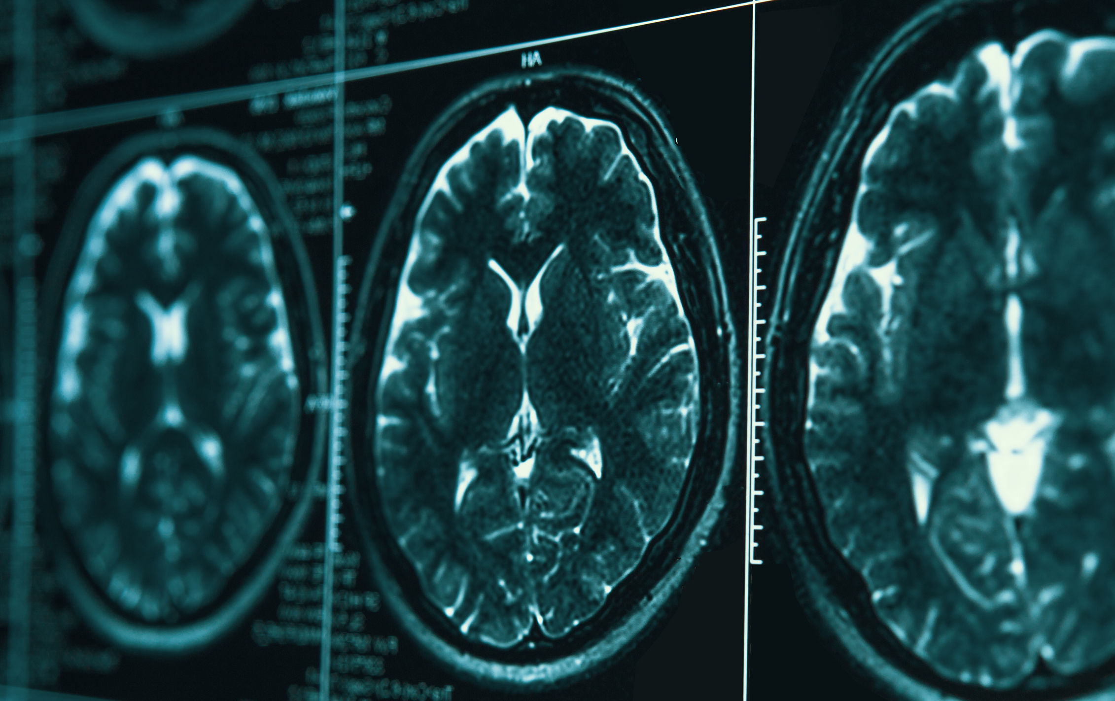
The team used deep learning to analyse almost 4,000 magnetic resonance (MR) scans from 715 patients, with the findings published in The New England Journal of Medicine AI. Rather than interpreting single images, the researchers employed a method known as temporal learning to train the AI on sequential post-surgery brain scans. This enabled the algorithm to detect subtle patterns of change over time, thereby improving its predictive ability.
Dr Benjamin Kann, corresponding author and a member of the Artificial Intelligence in Medicine (AIM) Programme at Mass General Brigham, said: “Many pediatric gliomas are curable with surgery alone, but when relapses occur, they can be devastating. It is very difficult to predict who may be at risk of recurrence, so patients undergo frequent follow-up with magnetic resonance (MR) imaging for many years, a process that can be stressful and burdensome for children and families. We need better tools to identify early which patients are at the highest risk of recurrence.”
The AI model demonstrated an accuracy rate of 75-89% in predicting recurrence within a year following treatment, compared with around 50% for traditional single-image analysis. The algorithm’s performance improved with more post-treatment timepoints, but plateaued after four to six scans.
Although the results were promising, the researchers acknowledged the need for further validation across additional clinical settings. Future trials are expected to explore whether AI-informed predictions could lead to changes in care, such as reducing scan frequency for low-risk patients or introducing earlier interventions for those at high risk.
“We have shown that AI is capable of effectively analysing and making predictions from multiple images, not just single scans,” said first author Divyanshu Tak, MS, of the AIM Program at Mass General Brigham and the Department of Radiation Oncology at the Brigham. “This technique may be applied in many settings where patients get serial, longitudinal imaging, and we’re excited to see what this project will inspire.”
The project received partial funding from the National Institutes of Health and was supported by cross-institutional data sharing to overcome the challenges associated with studying rare diseases like paediatric glioma.