
Adding colour to SEM imaging
The Scanning Electron Microscope (SEM) is widely used in various fields of industry and science and it allows users to see details 1,000 times smaller than a conventional microscope.
However, on the downside, images obtained by SEMs are two-dimensional and black and white (this is of course normal, as SEMs use electrons and not photons - or light rays - for visualisation). To make SEM images easier to interpret by the human eye, researchers spend hours ‘colorising’ their images manually before publishing them.
This week, the international scientific and industrial community will have at its disposal a new tool developed by Digital Surf - Mountains 7.3 software - which will enable colourisation of an object with just one click of the mouse.
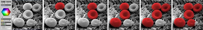
Behind this new tool, Mountains 7.3 software performs over 30 successive mathematical operations in order to distinguish different objects in the image. In the previous 7.2 version of the software, Digital Surf had already made it possible for SEM users to turn their 2D images into 3D models, an operation that requires hundreds of billions of instructions for a microprocessor.
Digital Surf is now the only company on the market to provide a comprehensive software solution for processing SEM images, as well as single all-in-one software solution for analysing data (surfaces and images) obtained by any type of microscope.
Digital Surf’s partners, including some of the leading SEM manufacturers, will be able to deliver this new version of the software to their users worldwide.
Product Spotlight
APV1111GVY
Panasonic

Panasonic PhotoMOS® Photovoltaic MOSFET High-Power Drivers
| SKU: | |
|---|---|
| Stock: | 3490 |
| Cost: | $3.95 |
